| Clinical Infection and Immunity, ISSN 2371-4972 print, 2371-4980 online, Open Access |
| Article copyright, the authors; Journal compilation copyright, Clin Infect Immun and Elmer Press Inc |
| Journal website https://cii.elmerpub.com |
Case Report
Volume 000, Number 000, July 2026, pages 000-000

Genital Tuberculosis in a Reproductive-Age Woman: A Diagnostic Challenge
Amina Etbera, b, Kaoutar Bahidaa, b, Soukaina Mouimana, Najia Zeraidia, Nisrine Benouichaa, Aziz Baidadaa
aGynecology-Obstetrics and Endoscopy Department, Maternity Souissi, University Hospital Center, IBN SINA, University Mohammed V, Rabat, Morocco
bCorresponding Authors: Amina Etber and Kaoutar Bahida, Gynecology-Obstetrics and Endoscopy Department, Maternity Souissi, University Hospital Center, IBN SINA, University Mohammed V, Rabat, Moroccoand
Manuscript submitted May 22, 2026, accepted June 8, 2026, published online July 30, 2026
Short title: GTB in a Reproductive-Age Woman
doi: https://doi.org/10.14740/cii514
| Abstract | ▴Top |
Genital tuberculosis (GTB) is a rare but serious form of extrapulmonary tuberculosis (EPTB) that significantly affects female reproductive health in endemic areas. It often presents as infertility or chronic pelvic pain, mimicking other gynecological disorders. A 31-year-old woman presented with fever, chronic pelvic pain, and secondary infertility. Pelvic ultrasound revealed bilateral tubo-ovarian masses, initially treated with antibiotics as a bacterial inflammatory disease, but GeneXpert on a sample of ultrasound-guided drainage confirmed Mycobacterium tuberculosis. She was started on a 6-month antitubercular therapy (HRZE regimen: isoniazid, rifampicin, pyrazinamide, and ethambutol) and is currently at the 3-month mark with noticeable clinical and sonographic improvement. GTB should be suspected in women with unexplained infertility, especially in tuberculosis-endemic countries. Early diagnosis and prompt antitubercular therapy are essential to prevent irreversible reproductive damage.
Keywords: Genital tuberculosis; Extrapulmonary tuberculosis; Infertility; Reproductive health
| Introduction | ▴Top |
Tuberculosis (TB) is a major global health concern; extrapulmonary forms account for 15–20% of all cases in immunocompetent individuals [1, 2]. Among these, genital tuberculosis (GTB) affects both male and female reproductive tracts but disproportionately impacts women of reproductive age [1, 2]. The spread occurs almost exclusively through hematogenous dissemination from a primary pulmonary site, with the female genital tract being particularly susceptible due to its rich blood supply [2, 3], although direct spread or lymphatic dissemination is also possible [4].
GTB accounts for 9–14% of extrapulmonary TB in women and remains an under recognized cause of infertility in developing countries [1, 2]. The fallopian tubes are involved in almost all cases (90–100%), with secondary spread to the endometrium (50–60%), ovaries (20–30%) [5], and cervix, leading to tubal damage and endometrial scarring, which are the principal mechanisms leading to GTB-related infertility [1, 3].
The clinical manifestations of GTB are not specific, they range from menstrual irregularities and chronic pelvic pain to primary or secondary infertility, while constitutional symptoms are often absent. This ambiguity of symptoms often results in misdiagnosis as endometriosis or pelvic inflammatory disease (PID), causing significant diagnostic delays [6, 7].
Advances in diagnostic methods such as polymerase chain reaction (PCR), histopathology, and laparoscopy have significantly improved the accuracy of the diagnosis, though no single modality is sufficient and a multimodal approach remains essential [7, 8]. Despite this progress, GTB is usually diagnosed late, after irreversible reproductive damage has already occurred [1, 3]. The following case highlights the diagnostic challenges in a reproductive-age woman with confirmed GTB, aiming to emphasize the importance of early suspicion in at-risk populations [6, 7].
This case illustrates that, though uncommon, GTB should always be considered in the differential diagnosis of chronic pelvic pain, recurrent tubo-ovarian abscesses unresponsive to conventional antibiotics or unexplained infertility in endemic regions [9]. A combination of histopathology, PCR or PCR-based methods, and laparoscopy improves diagnostic turnout. We also emphasize the importance of GeneXpert in comparison to the culture for a fast diagnosis of the TB leading to early antitubercular treatment, which is imperative to decrease morbidity, preserve reproductive potential, and prevent recurrence.
| Case Report | ▴Top |
Investigations
A 31-year-old woman, Gravida 1 Para 1, presented with intermittent lower abdominal pain accompanied by fever and a 10-year history of secondary infertility, even though the patient had no wish to conceive. Menstrual cycles were regular. The patient had no history of pulmonary TB or known contact exposure but reported previous ovarian cystectomy 7 years prior. On physical examination, lower abdominal tenderness was noted with a palpable mass in the left iliac fossa.
Pelvic ultrasound revealed bilateral tubo-ovarian abscesses, measuring approximately 13 × 10 cm on the left and 6 × 5 cm on the right. Pelvic computed tomography (CT) scan in non-contrast imaging confirmed bilateral complex adnexal masses with hyperdense content, showing no sign of pelvic peritonitis (Fig. 1), while post-contrast sequences (Fig. 2) showed characteristic peripheral rim enhancement. Laboratory analysis of the drained abscess samples using GeneXpert was positive for Mycobacterium tuberculosis DNA, confirming the tuberculous etiology. Chest X-ray showed no abnormalities, ruling out active pulmonary TB.
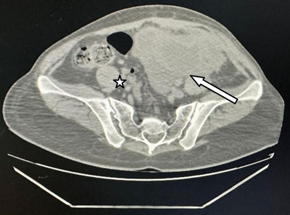 Click for large image | Figure 1. Non-contrast pelvic CT scan image of left (arrow) and right (star) tubo-ovarian abscess. CT: computed tomography. |
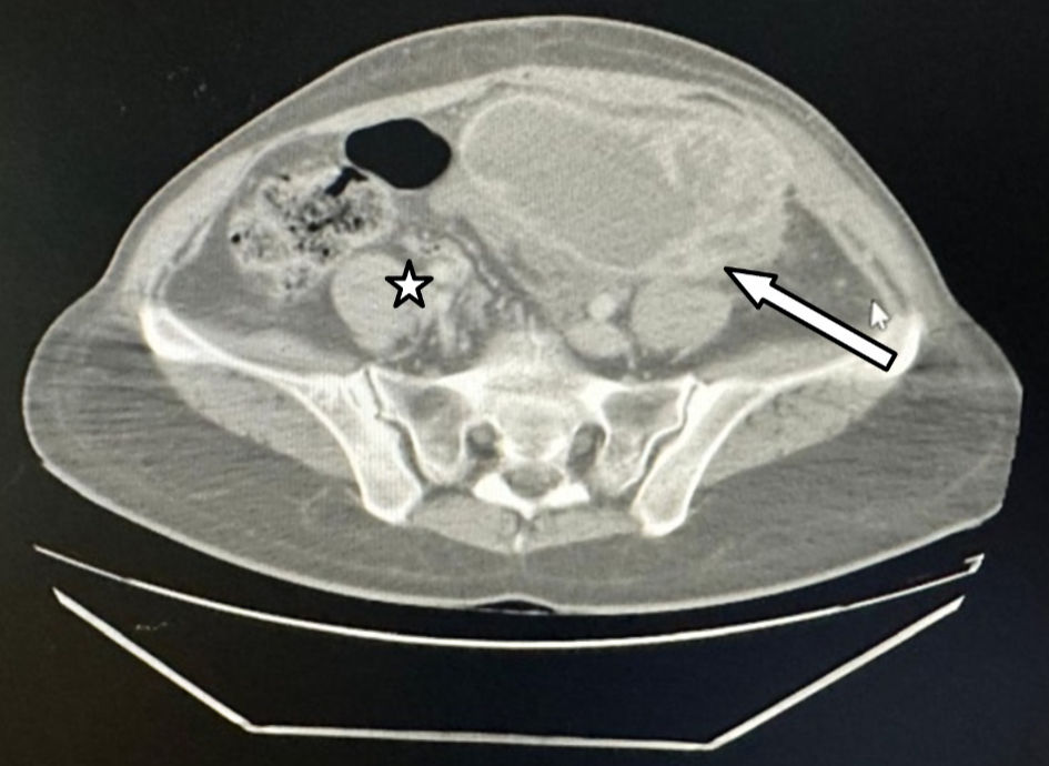 Click for large image | Figure 2. Contrast-enhanced pelvic CT scan image of left (arrow) and right (star) tubo-ovarian abscess. CT: computed tomography. |
Initial management
The patient received broad-spectrum antibiotic therapy with ceftriaxone, metronidazole, and doxycycline, without clinical or sonographic improvement.
Intervention
Ultrasound-guided drainage of the left tubo-ovarian abscess was performed since the mass was superficial and accessible. Samples were sent for microbiological analysis, which confirmed Mycobacterium tuberculosis complex DNA by GeneXpert.
Treatment
The patient was started on the World Health Organization (WHO)-recommended first-line antitubercular therapy (HRZE: isoniazid, rifampicin, pyrazinamide, and ethambutol) over a course of 6 months.
Follow-up and outcomes
Follow-up pelvic ultrasound at 1 month (Fig. 3) showed persistence of bilateral tubo-ovarian abscess with very slight size reduction in both the right and left adnexa. At 3 months (Fig. 4), the ultrasound check showed appreciable decrease in size and complexity of both abscesses, suggesting progressive resolution. By 6 months (Fig. 5), an almost complete radiological resolution of bilateral tubo-ovarian abscess was achieved without surgical intervention, matching a complete resolution of pain and pelvic mass.
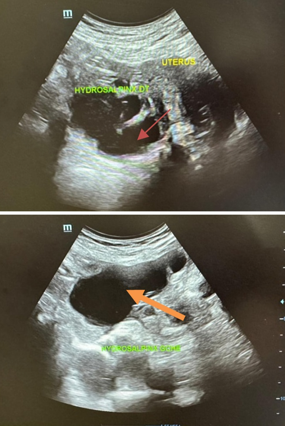 Click for large image | Figure 3. Ultrasound image of right (red arrow) and left (orange arrow) tubo-ovarian abscess after 1 month of antitubercular treatment. |
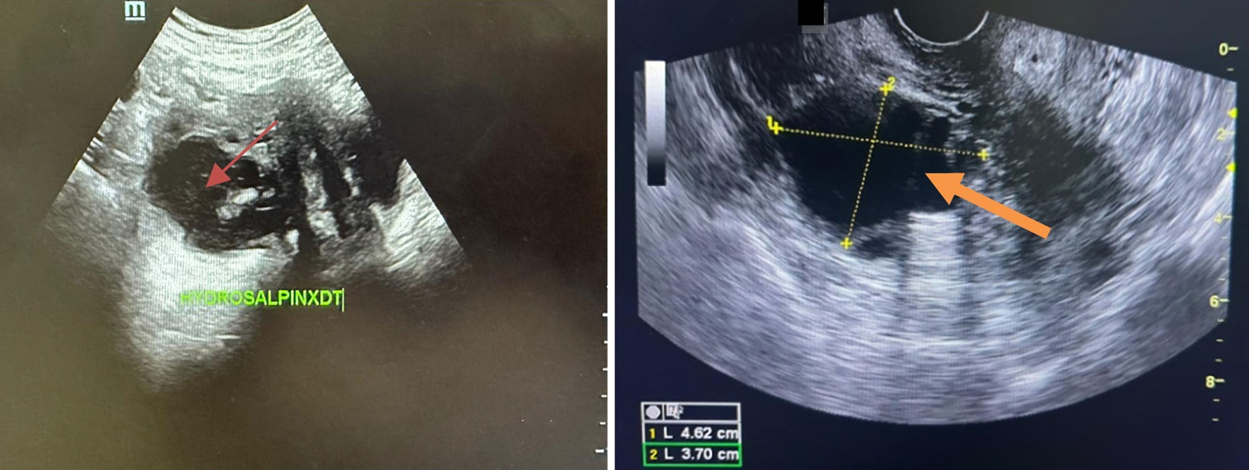 Click for large image | Figure 4. Ultrasound image of right (red arrow) and left (orange arrow) tubo-ovarian abscess after 3 months of antitubercular treatment. |
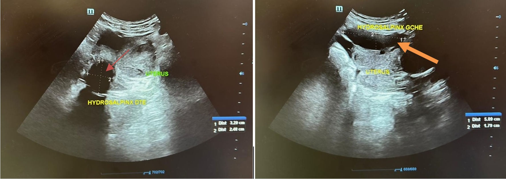 Click for large image | Figure 5. Ultrasound image of right (red arrow) and left (orange arrow) tubo-ovarian abscess after 6 months of antitubercular treatment. |
| Discussion | ▴Top |
The diagnostic modalities of GTB include histopathology, microbiological, molecular, and endoscopic techniques (Tables 1–3) [7, 10–12]. The histopathological identification of epithelioid granulomas with Langhans giant cells and caseous necrosis is a specific lesion of TB and remains diagnostic when present [10]. Ziehl-Neelsen staining for acid-fast bacilli, though specific when positive, carries low sensitivity. Culture on Lowenstein-Jensen medium or mycobacteria growth indicator tube (MGIT) remains the gold standard but is limited by slow growth of up to 8 weeks. Molecular methods, including PCR and GeneXpert MTB/RIF, offer high sensitivity and specificity with rapid turnaround, and are particularly valuable in paucibacillary samples and for simultaneous detection of rifampicin resistance (Table 1) [7, 10, 11]. Laparoscopy allows direct visualization of characteristic findings such as caseous nodules, beaded tubes, tubo-ovarian masses, and “violin-string” adhesions, and enables and facilitates targeted biopsy for histology and culture (Table 2) [12]. Hysteroscopy may also reveal intrauterine adhesions or a pale atrophic endometrium. Supportive tests, including erythrocyte sedimentation rate (ESR), C-reactive protein (CRP), Mantoux tuberculin skin testing, and interferon-gamma release assays (IGRAs), can assist in clinical decision-making but are neither specific nor confirmatory of genital involvement (Table 3).
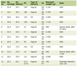 Click to view | Table 1. Microbiological and Molecular Diagnostic Tests for Genital Tuberculosis |
Click to view | Table 2. Endoscopic Diagnostic Modalities |
Click to view | Table 3. Supportive Diagnostic Tests |
The patient’s history of prior ovarian surgery may have contributed to diagnostic complexity, as postoperative adhesions or ovarian cysts can mimic features of chronic pelvic infections. In this case, the initial presentation with bilateral pyosalpinx raised suspicion for a conventional PID. Standard antibiotic therapy with ceftriaxone, metronidazole, and doxycycline, which typically covers common bacterial pathogens, failed to achieve clinical or sonographic improvement. This underscores the importance of considering atypical infections, such as TB, in persistent or recurrent tubo-ovarian abscesses.
Imaging studies played a critical role in assessing the extent of disease. Pelvic ultrasound identified bilateral tubo-ovarian abscess (left 13 × 10 cm; right 6 × 5 cm), while CT scan confirmed it and eliminated an associated pelvic peritonitis. The placement of an ultrasound-guided drain not only provided therapeutic benefit but also allowed for microbiological sampling, which ultimately confirmed the diagnosis through detection of Mycobacterium tuberculosis DNA using GeneXpert. The absence of pulmonary involvement on chest X-ray highlights that GTB can occur in isolation, and a high index of suspicion are necessary in women presenting with chronic pelvic masses or infertility.
Standard antitubercular therapy (HRZE: isoniazid, rifampicin, pyrazinamide, and ethambutol) remains the cornerstone of treatment for genital TB [13]. In this patient, 3 months of therapy resulted in a marked regression of the abscess on follow-up imaging, demonstrating the effectiveness of medical management. Surgical intervention is generally reserved for complications such as rupture, persistent abscess, or suspicion of malignancy; in this case, percutaneous drainage sufficed.
Despite microbiological and molecular advances, fertility outcomes remain poor, with conception rates of 10–20% post-therapy [5]. Assisted reproductive technologies, especially in vitro fertilization (IVF), offer the best chance of pregnancy in women with post-tubercular tubal damage [3].
While antitubercular therapy remains the cornerstone of treatment, surgical intervention plays a complementary role in selected cases, particularly in women seeking fertility restoration, which was not the case of our patient. In cases complicated by extensive tubo-ovarian abscesses or dense pelvic adhesions, laparoscopic adhesiolysis and tubal reconstructive procedures may help restore pelvic anatomy and improve reproductive potential [14, 15]. However, the success of tubal surgery in post-tubercular disease is significantly limited due to the extent of fibrosis and endosalpingeal destruction caused by the infection, resulting in poor spontaneous conception rates of 10–20% even following optimal medical and surgical management [16, 17]. Consequently, assisted reproductive technologies, particularly IVF, represent the most viable pathway to pregnancy in women with irreversible tubal damage, effectively bypassing the diseased fallopian tubes altogether [18]. In the present case, early diagnosis and prompt initiation of antitubercular therapy achieved significant abscess regression, preserving the opportunity for future fertility assessment and potentially reducing the need for more extensive surgical intervention. This underscores the importance of timely diagnosis, as earlier treatment may limit disease progression and maximize residual reproductive function [14, 16].
Conclusions
This case emphasizes several key clinical lessons. GTB should be suspected in women presenting with chronic pelvic pain, infertility, or recurrent tubo-ovarian abscesses unresponsive to standard antibiotic therapy. PCR-based methods, particularly GeneXpert, offer rapid and accurate diagnosis in settings where conventional cultures may be delayed or negative, and should be prioritized over culture when early treatment initiation is critical. A multimodal diagnostic approach combining histopathology, PCR, and laparoscopy optimizes diagnostic outcome. Finally, prompt initiation of antitubercular therapy, complemented by surgery when indicated, can lead to favorable outcomes including abscess resolution and preservation of reproductive potential in patients who wish to conceive.
Acknowledgments
None to declare.
Financial Disclosure
There are no funding sources to be declared.
Conflict of Interest
The authors declare that they have no competing interests.
Informed Consent
Written informed consent was obtained from the patient for publication of this case report and any accompanying images.
Author Contributions
Amina Etber, Kaoutar Bahida, and Soukaina Mouiman: study concept and design, data collection, data analysis and interpretation, and writing of the paper. Najia Zeraidi, Nisrine Benouicha, and Aziz Baidada: literature review, and supervision. The corresponding authors are the guarantors of submission.
Data Availability
The authors declare that data supporting the findings of this study are available within the article.
Abbreviations
AFB: acid-fast bacilli; CRP: C-reactive protein; EPTB: extrapulmonary tuberculosis; ESR: erythrocyte sedimentation rate; GTB: genital tuberculosis; HRZE: isoniazid, rifampicin, pyrazinamide, and ethambu-tol; IGRAs: interferon-gamma release assays; IVF: in vitro fertilization; MGIT: mycobacteria growth indicator tube; MTB: mycobacterium tuberculosis; PCR: polymerase chain reaction; PID: pelvic inflammatory disease; RIF: rifampicin resistance; TB: tuberculosis; WHO: World Health Organization; ZN: Ziehl-Neelsen
| References | ▴Top |
- Sharma JB, Sharma E, Sharma S, Dharmendra S. Female genital tuberculosis: Revisited. Indian J Med Res. 2018;148(Suppl):S71-S83.
doi pubmed - Grace GA, Devaleenal DB. Genital tuberculosis in women: overview and challenges. J Obstet Gynaecol Res. 2018;44(6):1027-1036.
- Kumar R, Singh M. Fertility outcomes following treatment of genital tuberculosis. Obstet Gynecol Int J. 2021;12(1):12-17.
- Singh N, Sumana G, Mittal S. Genital tuberculosis: a leading cause for infertility in women seeking assisted conception in North India. Arch Gynecol Obstet. 2008;278(4):325-327.
doi pubmed - Neema JP, et al. Reproductive outcome after antitubercular therapy in women with genital tuberculosis. J Hum Reprod Sci. 2020;13(2):123-129.
- Jindal N, et al. Female genital tuberculosis: Revisited. Indian J Tuberc. 2015;62(4):260-265.
- Banaei N, et al. Molecular diagnosis of extrapulmonary tuberculosis. J Clin Microbiol. 2016;54(7):1893-1899.
- World Health Organization. Global tuberculosis report 2022. WHO Press, Geneva. 2022. https://www.who.int/tb/publications/global_report.
- Singh N, Gupta A. Diagnostic challenges in female genital tuberculosis. Cureus. 2019;11(1):e3840.7.
- Kulshrestha V, et al. Genital tuberculosis and infertility: Experience from an endemic region. Arch Gynecol Obstet. 2016;293(5):1045-1052.
- Chakraborty S, et al. Utility of GeneXpert MTB/RIF assay in extrapulmonary specimens. Indian J Med Res. 2019;150(6):666-672.
- Arora R, Gupta N. Laparoscopic findings in female genital tuberculosis. Int J Gynecol Obstet. 2015;129(2):161-164.
- Sharma JB, Aggarwal P. Management of female genital tuberculosis and infertility: Current perspectives. Int J Womens Health. 2020;12:285-295.
- Sharma JB, Sneha J, Singh UB, et al. Comparative study of laparoscopic findings before and after antitubercular therapy in female genital tuberculosis with infertility. Journal of Minimally Invasive Gynecology. 2016;23(2):215-222.
- Gungorduk K, Ulker V, Sahbaz A, et al. Post-tuberculous hydrosalpinx and tubal factor infertility: surgical and assisted reproductive outcomes. Archives of Gynecology and Obstetrics. 2009;280(3):439-443.
- Namavar Jahromi B, Parsanezhad ME, Ghane-Shirazi R. Female genital tuberculosis and infertility. Int J Gynaecol Obstet. 2001;75(3):269-272.
doi pubmed - Tripathy SN, Tripathy SN. Infertility and pregnancy outcome in female genital tuberculosis. Int J Gynaecol Obstet. 2002;76(2):159-163.
doi pubmed - Dhaliwal LK, Gupta KR, Ganguly NK, Majumdar S. Genital tuberculosis and assisted reproduction. Journal of Assisted Reproduction and Genetics. 2008;25(7):303-306.
This article is distributed under the terms of the Creative Commons Attribution 4.0 International License (CC BY 4.0), which permits unrestricted use, distribution, and reproduction in any medium, including commercial use, provided the original work is properly cited.
Clinical Infection and Immunity is published by Elmer Press Inc.