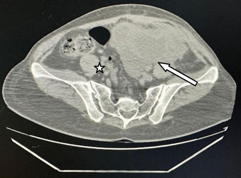
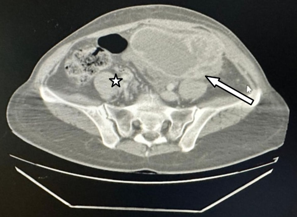
↓ Figure 1. Non-contrast pelvic CT scan image of left (arrow) and right (star) tubo-ovarian abscess. CT: computed tomography.
| Clinical Infection and Immunity, ISSN 2371-4972 print, 2371-4980 online, Open Access |
| Article copyright, the authors; Journal compilation copyright, Clin Infect Immun and Elmer Press Inc |
| Journal website https://cii.elmerpub.com |
Case Report
Volume 11, Number 1, August 2026, pages 6-11
Genital Tuberculosis in a Reproductive-Age Woman: A Diagnostic Challenge
Figures
Tables
| Modality | Key features | Limitations |
|---|---|---|
| PCR: polymerase chain reaction; ZN: Ziehl-Neelsen; MGIT: mycobacteria growth indicator tube. | ||
| Histopathology | Epithelioid granulomas with Langhans giant cells and caseous necrosis—diagnostic when present [10] | Requires tissue biopsy; not always available |
| ZN staining | Detects acid-fast bacilli (AFB); specific when positive | Low sensitivity; misses paucibacillary cases |
| Culture (gold standard) | Lowenstein-Jensen medium or MGIT liquid culture; highest diagnostic accuracy | Slow growth (up to 8 weeks); delays clinical decision-making |
| PCR | Detects Mycobacterium tuberculosis DNA with high sensitivity and specificity; particularly useful in paucibacillary samples [7] | Risk of contamination; variable sensitivity across sample types |
| GeneXpert MTB/RIF | Rapid detection of Mycobacterium tuberculosis and rifampicin resistance; increasingly used as a confirmatory test [11] | Limited sensitivity in extrapulmonary specimens |
| Modality | Key features | Limitations |
|---|---|---|
| TB: tuberculosis. | ||
| Laparoscopy [12] | Direct visualization of caseous nodules, beaded tubes, tubo-ovarian masses, and “violin-string” adhesions; allows targeted biopsy for histology and culture | Invasive; requires general anesthesia; operator-dependent |
| Hysteroscopy | May reveal intrauterine adhesions or pale, atrophic endometrium suggestive of endometrial TB | Findings are nonspecific; require endometrial sampling for confirmation |
| Test | Role | Limitations |
|---|---|---|
| GTB: genital tuberculosis; ESR: erythrocyte sedimentation rate; IGRAs: interferon-gamma release assays; TB: tuberculosis; CRP: C-reactive protein; BCG: bacillus Calmette-Guerin. | ||
| ESR/CRP | Often elevated in active GTB; useful for monitoring treatment response | Nonspecific; elevated in many inflammatory conditions |
| Tuberculin skin test (Mantoux) | May support diagnosis in the right clinical context | Not specific for genital involvement; false positives in BCG-vaccinated individuals |
| IGRAs | Useful for diagnosing latent TB infection | Not confirmatory for genital TB; cannot distinguish active from latent disease |