| Clinical Infection and Immunity, ISSN 2371-4972 print, 2371-4980 online, Open Access |
| Article copyright, the authors; Journal compilation copyright, Clin Infect Immun and Elmer Press Inc |
| Journal website https://cii.elmerpub.com |
Case Report
Volume 000, Number 000, July 2026, pages 000-000

Genital Herpes Zoster Infection
Ellen Carona, b, Kathleen Wuytsa
aDepartment of Obstetrics and Gynecology, General Hospital Lier, 2500 Lier, Belgium
bCorresponding Author: Ellen Caron, Department of Obstetrics and Gynecology, General Hospital Lier, 2500 Lier, Belgium
Manuscript submitted May 26, 2026, accepted July 2, 2026, published online July 30, 2026
Short title: Genital Herpes Zoster Infection
doi: https://doi.org/10.14740/cii515
| Abstract | ▴Top |
A 75-year-old woman presented at the emergency room with pain at the left labia and appearance of edema, erythema and vesicular lesions. The woman had chronic obstructive pulmonary disease (COPD) and was receiving methylprednisolone therapy. Clinical findings and medical history suggested an infection of herpes simplex virus (HSV); however, polymerase chain reaction (PCR) swab of the lesions was negative for HSV and positive for Pseudomonas aeruginosa. Ceftazidime was started, and given the clinical picture, acyclovir was added. Despite treatment, the patient’s symptoms worsened, requiring surgical debridement. Eventually, the diagnosis of herpes zoster infection was made. The literature shows that 2.9% of diagnoses with genital HSV are misdiagnosed and represent varicella-zoster virus (VZV). Given the rare presentation of genital herpes zoster (HZ), this is a little-described topic in the literature.
Keywords: Herpes zoster; Varicella zoster virus; Herpes genitalis; Genital lesions
| Introduction | ▴Top |
Varicella zoster virus is a double-stranded DNA virus which can cause chickenpox in the primary infection [1]. Afterwards, the virus remains latent in the posterior dorsal root ganglion [2]. Reactivation occurs usually decades after the initial infection and causes herpes zoster (HZ) or shingles [3]. Most often, involvement of the thoracic and lumbar nerves (dermatomes T3–L3) occurs. Involvement of the sacral plexus is seen only in 4–8% of cases, with even rarer affection of the vulvar area in only 2%. Infection arises mainly in immunocompromised or human immunodeficiency virus (HIV)-positive patients [2].
Early symptoms include pain, edema and erythema in the involved dermatome, typically unilateral. Later on, a maculopapular rash and development of vesicles appear, with crusting over time. In addition, fever, headache and enlarged lymph nodes may be present [2, 4].
Diagnosis should be made via sampling of open lesions, on which polymerase chain reaction (PCR) assay can be performed [5]. This is the quickest and most sensitive test [3]. Approximately 2.9% of initial diagnoses of genital herpes simplex virus (HSV) infection are eventually a varicella-zoster virus (VZV) infection [5]. Treatment should ideally be started within 72 h of the onset of the first symptoms. Therapy options include acyclovir, valaciclovir and famciclovir [6–8].
| Case Report | ▴Top |
This case describes a 75-year-old female patient who presented to the emergency room with progressively increasing edema and pain at the left labia. Gynecologic examination revealed erythematous swelling of the left labium majus and labium minus. The same region showed vesicular to verrucous lesions, extending to the buttock seam on the left side, in dermatomes S4–S5 (Fig. 1). No lymphadenopathy was present. Further anamnesis revealed that the patient had a regular sexual partner for several years and experienced normal vaginal discharge. The family doctor had already prescribed amoxicillin–clavulanic acid 875 mg three times daily orally without improvement in symptoms. Given the differential diagnosis at that time, including herpes simplex and genital warts, vaginal and lesion swabs were taken for PCR testing for herpes simplex.
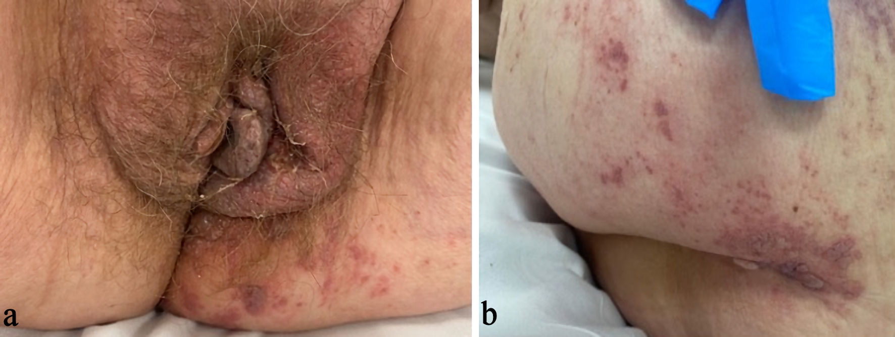 Click for large image | Figure 1. Clinical evolution of the infection. Day 0: Status at first presentation to the emergency department. (a) Edematous swelling of the left labia, with several blisters extending to the buttock. (b) Reddish raised areas on the left buttock, some with a verrucous appearance. |
After emergency presentation, acyclovir 800 mg three times daily orally was initiated, and amoxicillin–clavulanic acid was continued. Given the suspicion of bacterial superinfection, local Isobetadine application was recommended. Swabs were negative for herpes simplex; however, bacterial superinfection with Pseudomonas aeruginosa was identified. Antibiotic therapy was changed to intravenous ceftazidime 2 g three times daily to treat this superinfection, together with locally applied diluted vinegar. Despite treatment, clinical progression of symptoms and lesions occurred (Figs. 2, 3).
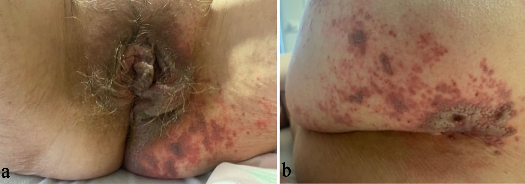 Click for large image | Figure 2. (a, b) Day 4: Increase in the number and extent of lesions with the appearance of a grayish wound coating. |
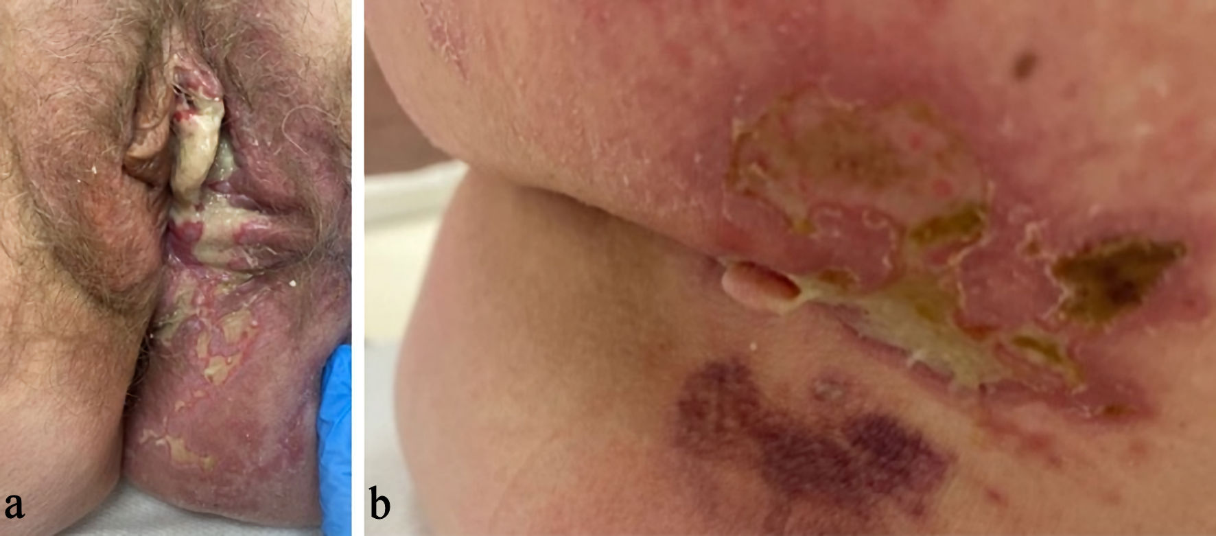 Click for large image | Figure 3. Day 14. (a) New purulent discharge from the left vulvar lesions. (b) Improved healing of the lesions on the left buttock. |
To exclude atypia, vulvoscopy with biopsy was performed. Vulvoscopy revealed a large open wound extending from the entire left labia majora and minora to the buttock seam at the level of the sacrum, with wound slough. Biopsies were obtained from the coccyx, left buttock, and left labium minus. Due to further extension of the lesions with the development of purulent discharge, the patient was transferred to a university hospital. Surgical debridement and drainage of the lesions were performed, new swabs were obtained, and intravenous acyclovir (800 mg three times daily) and piperacillin/tazobactam (4 g four times daily) were started. Swab testing was positive for VZV. The patient was retransferred after clinical improvement, although she developed new neuropathic pain in the S1–S2 dermatomes, for which gabapentin was initiated. Further improvement in symptoms and clinical appearance was observed, and the patient was discharged from hospital. Examination 2 weeks later demonstrated a persistent, well-controlled infection (Fig. 4).
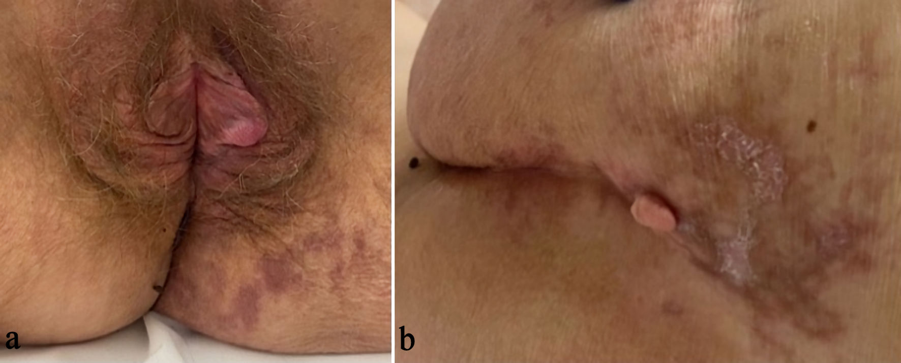 Click for large image | Figure 4. (a, b) Day 49: healing of all lesions. |
| Discussion | ▴Top |
HZ or shingles is caused by the reactivation of the VZV. This virus causes chickenpox at primary infection and subsequently travels to the dorsal ganglia where it remains latent [1]. Reactivation occurs mainly in immunocompromised individuals and occurs with a typical presentation of unilateral vesicular lesions spread according to a particular dermatome. It mainly occurs in T3–L3; however, in rarer cases (4–8%), it presents in the sacral plexus and even rarer in the vulvar region [2].
Given its rare presentation in the genital area, HZ is often under- or misdiagnosed. This was observed in a study by Birch et al, where 6,210 genital samples were analyzed by multiplex PCR testing. A total of 2,225 samples were found positive, 36% of them for HSV-1, 61% for HSV-2 and 2.9% for VZV. Of these genital VZV infections, most were initially clinically assessed as HSV [5].
In a 1-year investigation, Granato et al examined 2,113 cutaneous and mucocutaneous samples for HSV-1, HSV-2 and VZV. Approximately 6% of the samples were found positive for VZV, of which 11.1% unexpectedly originated from the genital area (in both males and females) [9].
Risk factors leading to reactivation of the VZV include aging, HIV infection, family history, immunocompromised state [10], and use of immunosuppressants, with an increased risk observed among patients receiving tumor necrosis factor-alpha inhibitors [2]. Chronic conditions such as diabetes mellitus, cardiovascular and respiratory diseases also carry an elevated risk of reactivation [11].
Various methods for diagnostic testing for varicella zoster have been described. Samples should ideally be taken from open lesions, which can be used for virus culture, direct fluorescent antibody testing, or PCR to detect viral DNA [2]. Although virus culture is a sensitive test with a positivity rate of 80–99%, it requires more time [3]. Diagnosis via immunofluorescence testing is less sensitive than virus culture and PCR testing. The latter has proven to be the fastest and most sensitive method and is therefore the preferred means of diagnosis [2, 3]. Serological testing may also be performed, with detection of immunoglobulin (Ig)G and IgM for HSV and VZV for additional information [12].
Treatment is recommended to be initiated within 72 h of the onset of symptoms, ideally before the appearance of lesions. This reduces the severity and duration of symptoms and moreover reduces the risk of complications such as post-herpetic neuralgia [13]. Higher daily doses should be given for VZV than for HSV. In this case, treatment with acyclovir 800 mg orally three times daily did not result in sufficient improvement. Rather, the frequency should be increased to five times daily when treating HZ [4]. The antiviral drug of first choice is still controversial in the treatment of HSV. A randomized, double-blind study published in 2001 described the similar effect of oral famciclovir and acyclovir in immunocompromised patients. A trend towards a shorter acute phase with famciclovir compared to acyclovir was observed, but this difference was not significant [6]. Ono et al confirmed in their study the reduced duration of acute pain with famciclovir compared to valaciclovir [7]. In contrast, a review study by Patil et al described only the usefulness of famciclovir in cases of acyclovir resistance, and not as a primary treatment option [8]. Given the higher bioavailability of famciclovir and valaciclovir compared to acyclovir, the reduction in daily intake frequency provides an advantage in terms of compliance [6].
The correct diagnosis is necessary not only for determining the dosage for treatment, but also for the psychological factors involved. Since HSV is transmitted through sexual contact and HZ develops after reactivation of VZV, the correct diagnosis is an important factor from a mental and relational point of view. On the other hand, the possible transmission of the virus in open lesions to non-immune persons must also be taken into account. If lesions are present in the genital area, the virus can also be transmitted through sexual contact [1].
The most common complication of HZ is postherpetic neuralgia (PHN), which is seen in 5% to 15% of patients with HZ [14]. It occurs approximately 4 weeks after the onset of the initial HZ symptoms and can persist for weeks up to months [8, 14]. An increased risk of PHN occurs with higher age, decreased immunity, and severe acute pain in HZ. Therefore, it is recommended to start antiviral and pain medication as quickly as possible [14, 15]. Women also appear to be more likely to develop PHN than men [13]. Local treatment of PHN consists of lidocaine 2% gel, capsaicin 0.05% ointment, or capsaicin 8% patches [14, 15]. Systemic treatment includes nonsteroidal anti-inflammatory drugs (NSAIDs) or opioids. Tricyclic antidepressants, such as amitriptyline or nortriptyline, have been used perviously; however, given the side effects like anticholinergic effects, antiepileptics are now preferred (e.g., gabapentin or pregabalin) [13, 15].
Conclusions
This case once again highlights the difficulty of diagnosing genital HZ, considering its rarity and similarities to herpes simplex infection. Caution to HZ should always be applied in cases of genital vesicular lesions with an unilateral pattern in immunocompromised patients. PCR testing of genital lesions for HSV1, HSV2, as well as VZV should become the new standard. Correct diagnosis is necessary for the early initiation of treatment, preferably within 72 h, and with a higher dose for VZV than HSV. The most common complication of VZV is PHN, with antiepileptic medicines such as gabapentin as primary treatment.
Acknowledgments
None to declare.
Financial Disclosure
This research received no external funding.
Conflict of Interest
None to declare.
Informed Consent
Informed consent was obtained.
Author Contributions
Conceptualization: EC and KW. Methodology: EC and KW. Writing—original draft preparation: EC and KW. Writing—review and editing: EC and KW. Visualization: EC and KW. Both authors have read and agreed to the published version of the manuscript.
Data Availability
Data supporting the findings of this study are available within the article.
Abbreviations
HSV: herpes simplex virus; VZV: varicella-zoster virus; HZ: herpes zoster; PHN: postherpetic neuralgia
| References | ▴Top |
- Granato PA. Genital zoster infection: the great imposter. Clinical Microbiology Newsletter. 2018;40(19):157-162.
- Martins MM, Ferreira P, Maciel R, Costa C. Vulvar herpes zoster infection: a rare and challenging diagnosis. BMJ Case Rep. 2021;14(12).
doi pubmed - Kalantzi S, Alexakis C, Anagnostaki ME, Anagnostaki I, Zacharis K. Genital herpes zoster: a report of a rare case. Cureus. 2024;16(10):e72290.
doi pubmed - Karalok H, et al. Herpes zoster infection of the vulva. Turkiye Klinikleri J Gynecol Obst. 2007;17(5):393-395.
- Birch CJ, Druce JD, Catton MC, MacGregor L, Read T. Detection of varicella zoster virus in genital specimens using a multiplex polymerase chain reaction. Sex Transm Infect. 2003;79(4):298-300.
doi pubmed - Tyring S, Belanger R, Bezwoda W, Ljungman P, Boon R, Saltzman RL, Colaborative Famciclovir Immunocompromised Study G. A randomized, double-blind trial of famciclovir versus acyclovir for the treatment of localized dermatomal herpes zoster in immunocompromised patients. Cancer Invest. 2001;19(1):13-22.
doi pubmed - Ono F, Yasumoto S, Furumura M, Hamada T, Ishii N, Gyotoku T, Higuchi M, et al. Comparison between famciclovir and valacyclovir for acute pain in adult Japanese immunocompetent patients with herpes zoster. J Dermatol. 2012;39(11):902-908.
doi pubmed - Patil A, Goldust M, Wollina U. Herpes zoster: a review of clinical manifestations and management. Viruses. 2022;14(2).
doi pubmed - Granato PA, DeGilio MA, Wilson EM. The unexpected detection of varicella-zoster virus in genital specimens using the Lyra Direct HSV 1+2/VZV Assay. J Clin Virol. 2016;84:87-89.
doi pubmed - Gruver C, Guthmiller KB. Postherpetic neuralgia. In: StatPearls. Treasure Island (FL) ineligible companies. 2026.
pubmed - Oleszko M, Zapolnik P, Kmiecik W, Czajka H. Herpes zoster: risk factors for occurrence, complications, and recurrence with a focus on immunocompromised patients. Diseases. 2025;13(3).
doi pubmed - Philip KE, Goodman A, Pallawela SN, Sathia L, Webster DP. A not so simplex case of genital herpes. BMJ Case Rep. 2013;2013:bcr2013009993.
doi pubmed - Koshy E, Mengting L, Kumar H, Jianbo W. Epidemiology, treatment and prevention of herpes zoster: a comprehensive review. Indian J Dermatol Venereol Leprol. 2018;84(3):251-262.
doi pubmed - Aggarwal A, Suresh V, Gupta B, Sonthalia S. Post-herpetic neuralgia: a systematic review of current interventional pain management strategies. J Cutan Aesthet Surg. 2020;13(4):265-274.
doi pubmed - Lim DZJ, Tey HL, Salada BMA, Oon JEL, Seah ED, Chandran NS, Pan JY. Herpes zoster and post-herpetic neuralgia-diagnosis, treatment, and vaccination strategies. Pathogens. 2024;13(7).
doi pubmed
This article is distributed under the terms of the Creative Commons Attribution 4.0 International License (CC BY 4.0), which permits unrestricted use, distribution, and reproduction in any medium, including commercial use, provided the original work is properly cited.
Clinical Infection and Immunity is published by Elmer Press Inc.